#Exposure x ray skin
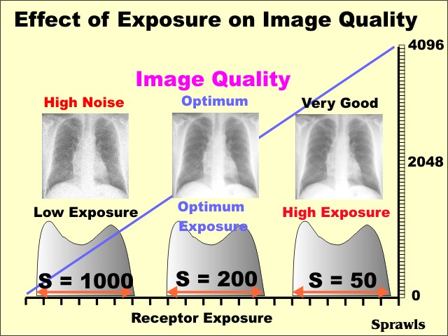
Peak skin dose values are determined from (1) measuring the X-ray tube output (air kerma per mAs as a function of kVp at fixed filtration) at a fixed distance and determining incident air kerma to the patient based upon knowledge of the kVp and mAs and corrections for variation in distance (2) using a kerma area product (KAP) or reference dose device positioned at the X-ray tube collimator that physically measures x-radiation emanating from the focal spot, to generate a dose area product or a reference point dose, either of which can provide an estimate of the incident dose to the patient. Peak skin dose and effective dose are the most common. There are several ways to estimate the dose to the patient. Dose to the patient is dependent upon many factors, including the parts of the body being examined, presence or lack of radiation-sensitive organs being exposed to X-rays, the area of the X-ray beam (determined by collimation) irradiating the patient, the output of the X-ray tube as a function of kVp, tube current, exposure time and beam filtration. Although these are important values in providing feedback in regard to achieving optimal images at the lowest dose, the EI and DI don’t directly indicate patient dose. The goals of each group were recently completed, with the resultant IEC standard 62494-1 “exposure index of digital X-ray imaging systems,” which provides a unified method to generate an exposure index value, requires users to input a target exposure index (EI T) value for each exam, and indicates a deviation index (DI) value that gives feedback to the technologist regarding technique and image quality based on signal-to-noise ratio. An effort to standardize the EI for all digital radiography detector systems was initiated by the International Electrotechnical Commission (IEC) and the American Association of Physicists in Medicine (AAPM) with participation by diagnostic physicists from around the world and representatives from many digital radiography manufacturers. Currently, because of the many proprietary and distinctly different ways of determining and reporting the EI by the manufacturers of digital radiography equipment, there has been confusion regarding use of this value from the technologist, radiologist and physicist communities. The exposure index (EI) in digital radiography has been used to indicate the relative speed and sensitivity of the digital receptor to incident X-rays and, ideally, to provide feedback to the technologist regarding the proper radiographic techniques for a specific exam that achieves an optimal image in terms of appropriate quality and corresponding low dose to the patient. The Alliance for Radiation Safety in Pediatric Imaging, in its role as a benefactor of and advocate for the pediatric patient, is using the Image Gently campaign to disseminate information regarding the exposure index standard for digital radiography so that these benefits can be achieved in a rapid and effective manner. Radiologists will benefit from standardized terminology, and institutions and clinics will be able to compare exposure index values with others through a national dose index registry database now under development.

However, the use of the standardized exposure index and its associated target exposure index and deviation index values will likely lead to improved technologist performance in terms of uniformity and use of optimized radiographic techniques, leading to safer care of children needing radiographic examinations. As explained, the exposure index does not indicate patient dose but rather a linearly proportional estimate of the incident radiation exposure to the detector. Developed concurrently by the International Electrotechnical Commission and the American Association of Physicists in Medicine in cooperation with digital radiography system manufacturers, the index has been implemented as an international standard. Fortunately, a new exposure index of digital X-ray imaging systems has been implemented.

Unfortunately, there are as many exposure index values and methods as there are manufacturers, and in an environment with multiple vendors and a need to share data across institutions and dose registry databases, the situation is complicated.

The exposure index is currently a method by which digital radiography manufacturers provide feedback to the technologist regarding the estimated exposure on the detector, as a surrogate for image signal-to-noise ratio and an indirect indication of digital image quality.


 0 kommentar(er)
